What is Distal Radioulnar Joint (DRUJ) Instability?
Distal radioulnar joint instability is the abnormal orientation or movement of the radius and ulna bones at the wrist in relation to one another. Injury to the tendons, ligaments and/or muscles stabilizing the joint may cause partial or complete dislocation.
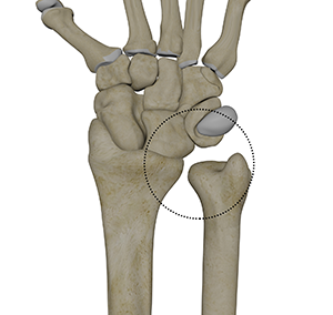
Anatomy
The articular surface of the radius bone referred to as the ulnar notch and the head of the ulna bone form the distal radioulnar joint. A complex structure called the triangular fibrocartilage complex (TFCC) contributes to wrist stability.
Symptoms
A deformed wrist may be a sign of DRUJ instability. Other symptoms include:
- Wrist pain
- Inflammation
- Weakness
- Decreased range of motion
Risk Factors
The common risk factors include:
- Trauma
- Fall on an outstretched hand
- Congenital bone defects
- Sports such as baseball, racquetball or tennis
Diagnosis
Your doctor will assess your symptoms, take your medical history, and perform a physical exam. Imaging tests such as X-ray, MRI or CT-scans may be ordered.
Treatment
Non-surgical Treatment
Non-surgical treatment options include:
- Rest and activity modification
- Immobilization with a splint or cast
- Physical therapy
Surgical Treatment
Surgery is recommended by your doctor if you do not respond to non-surgical treatment options, and involves the following:
- The surgery may be performed under general or local anesthesia.
- A few small incisions are made at the wrist near the radioulnar joint.
- An arthroscope, a small, fiber-optic instrument consisting of a lens, light source, and video camera, is inserted. The camera projects images of the inside of the joint onto a large monitor, allowing your surgeon to look for any damage, assess the type of injury and repair the problem.
- Debridement, or cleaning out the damaged tissue, is performed by your surgeon.
- A separate larger incision may need to be made to gain better access to the ulnar side of wrist
- Sutures may be used to re-attach the separated ligament back to the bone
The incision is closed and a bandage is applied.
Related Topics
- Wrist Fracture
- Fractures of the Hand and Fingers
- Wrist Sprain
- Flexor Tendon Injuries
- Distal Radioulnar Joint (DRUJ) Arthritis
- Ulnar Nerve Compression in Guyon's Canal
- Scaphoid Facture
- Industrial Hand Trauma
- Distal Radius Osteotomy to Correct Mal-Union (Crooked Painful Wrist)
- Distal Intersection Syndrome
- Distal Biceps Avulsion
- Adult Forearm Fractures
- Arthritis of the Hand and Wrist
- Arthritis of the Thumb
- Ganglion Cyst
- Boutonniere Deformity
- Carpal Tunnel Syndrome
- De Quervain's Tendinosis
- Dupuytren's Contracture
- Hand Pain
- Hand Infections
- Trapeziometacarpal (TMC) Arthritis
- Wrist Injuries
- Wrist Tumors
- Boxer's Fracture
- Swan Neck Deformity
- Carpal Instability
- Bennett's Fracture
- Kienbock's Disease
- Scapholunate Dissociation
- Triscaphoid Joint Arthritis
- Ulnar Carpal Impaction
- Triangular Fibrocartilage Complex Injury (TFCC)
- Guyon's Canal Syndrome
- Hand Masses
- Distal Radioulnar Joint (DRUJ) Instability
- Work Related Hand Injuries
- Wrist Ligament Tear and Instability
- Metacarpophalangeal Joint Arthritis